Retinal Photographs
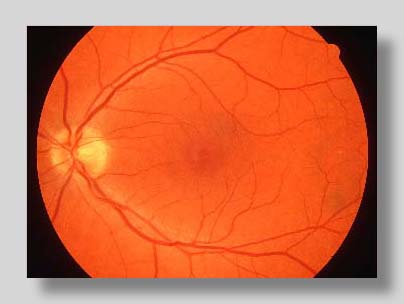
This photograph of a normal retina is taken through a dilated pupil with a special instrument called a fundus camera. The small, yellowish circle is the head of the optic nerve through which signals from the eye are sent to the brain. This optic disc measures about 1 1/2 millimeters in diameter. Retinal blood vessels enter the eye at the optic disc, and form a arcuate pattern around the macula which is responsible for central vision. At the center of the macula lies the tiny fovea, which produces the fine visual detail necessary for reading.
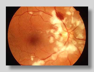
This photograph of a patient with Purtscher's Retinopathy demonstrates marked differences when compared with the normal retina above. Purtscher's Retinopathy is a disorder of the retina following severe trauma like an automobile accident. The blood flow is disrupted, causing bleeding or hemorrhage. Lack of blood supply to parts of the retina causes the white, fluffy appearance of cotton wool spots. |


