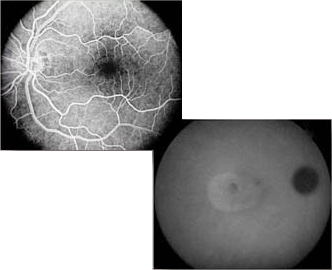
Angiography
The word Angiography comes from the Greek angeion, "vessel" and graphien, "to write or record". Angiography is the imaging of vessels, and the resulting pictures are angiograms. Angiography of the retina of the eye requires the injection of a small amount of dye into a vein in the patient's arm. The dye travels through the blood stream and is photographed using special cameras and colors of light as it travels through the vessels of retina. Fluorescein Angiography is performed with a dye called Sodium Fluorescein. When illuminated with a blue light, the fluorescein dye glows or fluoresces in yellow-green. Special filters in the camera allow only the fluorescent yellow-green light to be photographed, producing high contrast images of the retinal vessels. Fluorescein angiography can be performed using either standard black and white photographic film or using digital cameras. Indocyanine Green (ICG) dye is used in conjunction with infrared "light" for angiography in very special cases where fluorescein angiography proves inadequate. Because infrared energy is not in the visible spectrum and can not be imaged well with photographic film, high-sensitivity digital cameras are used for ICG angiography. |

