|
|
 |
Fundamentals of Fluorescein Angiography
Introduction
Timothy J. Bennett, CRA FOPS
Department of Ophthalmology
Penn State University
Hershey, Pennsylvania
From Current Concepts in Ophthalmology, Volume 9, 2001
A Publication of the Pennsylvania Academy of Ophthalmology
Used by permission
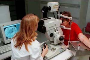
Fundus Camera with a Digital Capture System
Since the early 1960’s, ophthalmologists have relied on fluorescein angiography as an important tool in the understanding, diagnosis and treatment of retinal disorders. This diagnostic procedure utilizes a specialized fundus camera to capture rapid-sequence photographs of the retinal vasculature following an intravenous injection of fluorescein sodium. Fluorescein angiography facilitates the in-vivo study of the retinal circulation and is particularly useful in the management of diabetic retinopathy and macular degeneration, two common causes of blindness.
|
|
|