Fundamentals of Fluorescein Angiography
Indications and Uses
Timothy J. Bennett, CRA FOPS
Department of Ophthalmology
Penn State University
Hershey, Pennsylvania
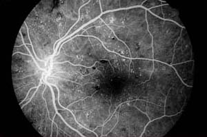
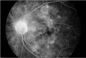
The most common uses of fluorescein angiography are in retinal or choroidal vascular diseases such as diabetic retinopathy, macular degeneration, hypertensive retinopathy and vascular occlusions. For the most part, these are clinical diagnoses. The angiogram is used to determine the extent of damage, to develop a treatment plan or to monitor the results of treatment. In diabetic retinopathy the angiogram is useful in identifying the extent of ischemia, the location of microaneurysms, the presence of neovascularization and the extent of macular edema. In macular degeneration, angiography is useful in identifying the presence and location of subretinal neovascularization. Post-treatment angiograms are used to check the efficacy of laser treatment.
Nonproliferative Diabetic Retinopathy

Venous Phase Photograph
Retinal Hemorrhages and Microaneurysms
|

Late Phase Photograph
Macular Edema
|
Macular Degeneration

Juxtafoveal Choroidal New Vessel Membrane
Other uses include degenerative and inflammatory conditions. Some of these conditions exhibit characteristic staining patterns, which can confirm the diagnosis. Stargardt's Disease is an example, exhibiting a silent choroid and a central bulls-eye staining pattern in the macula.
Angiography has long played a role in advancing the understanding of retinal vascular disorders and potential treatment modalities. A number of multicenter clinical trials utilize fluorescein angiography in investigating new treatment options in diabetic retinopathy and macular degeneration.
Photodynamic therapy (PDT) is a treatment option for macular degeneration that relies on pre-treatment angiograms to determine the location and size of the lesion to be treated. Digital imaging software is used to measure the actual size of the lesion and calculate the appropriate spot size of the PDT laser.

Digital Image of a Choroidal New Vessel Membrane
with Area Measurement for Photodynamic Therapy
|
Common Diagnostic uses and
Indications for Fluorescein Angiography
Diabetic retinopathy
Age related macular degeneration
Subretinal neovascular membrane
Central retinal vein occlusion
Branch retinal vein occlusion
Central serous chorioretinopathy
Cystoid macular edema
Hypertensive retinopathy
Central retinal artery occlusion
Branch retinal artery occlusion
Retinal arterial macroaneurysms
Pattern dystrophies of the retinal pigment epithelium
Choroidal tumors
Chorioretinal inflammatory conditions
Hereditary retinal dystrophies
|
|


