Fundamentals of Fluorescein Angiography
Phases of the Normal Angiogram
Timothy J. Bennett, CRA FOPS
Department of Ophthalmology
Penn State University
Hershey, Pennsylvania
Early Phase
The early phase of the angiogram can be broken down into distinct circulation phases that are useful in planning a photographic strategy and interpreting the results:
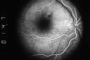
1. Choroidal flush. In a normal patient, the dye appears first in the choroid in 10-12 seconds. The major choroidal vessels are impermeable to fluorescein, but the choriocapillaris leaks fluorescein dye freely into the extravascular space. There is usually little detail in the choroidal flush as the retinal pigment epithelium (RPE) acts as an irregular filter that partially obscures the view of the choroid. If a cilioretinal artery is present, this fills along with the choroidal flush as both are fed by the short posterior ciliary artery.
2. Arterial phase. The retinal arterioles typically fill a second or two after the choroid. Complete filling of the retinal capillary bed follows the arterial phase and the retinal veins begin to exhibit laminar filling.

3. Venous phase. Complete filling of the veins occurs over the next ten seconds with maximum vessel fluorescence occurring within 30-35 seconds after injection.

Mid Phase
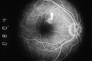
Also known as the recirculation phase, this occurs about 2 to 4 minutes after injection. The veins and arteries remain roughly equal in brightness. The intensity of fluorescence diminishes slowly during this phase as a significant amount of dye is removed from the bloodstream on the first pass through the kidneys.
Late Phase
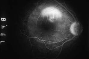
The late phase (or elimination phase) demonstrates the gradual elimination of dye from the retinal and choroidal vasculature. Staining of the optic disc is a normal finding in late phase images. Any other areas of late hyperfluorescence suggest the presence of an abnormality.

|