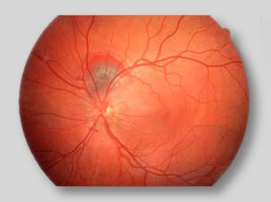
Ocular Fundus Imaging
Fundus is the bottom or base of anything. In medicine, it is a general term for the inner lining of a hollow organ. The ocular fundus is the inner lining of the eye made up of the Sensory Retina, the Retinal Pigment Epithelium, Bruch's Membrane, and the Choroid. Have you ever taken a fundus photograph? If you have taken photographs of your family or friends using a flash attachment on your camera and noticed that they have "red eyes", then you have! Photographic red eye is nothing more than a reflection of light off of the fundus of the eye. When performing ophthalmic fundus photography for diagnostic purposes, the pupil is dialted with eye drops and a special camera called a fundus camera is used to focus on the fundus. The resulting images can be spectacular, showing the optic nerve through which visual "signals" are transmitted to the brain and the retinal vessels which supply nutrition and oxygen to the tissue set against the red-orange color of the pigment epithelium. Color images like the one above provide documentation of the ocular fundus. Monochromatic, or "single color" images using colored filters in the light path of the fundus camera with black and white film can be used to isolate and study very specific parts of the retinal layer. The Nerve Fibre Layer of the retina can be photographed in this way. |

