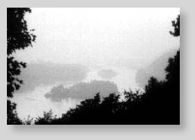
Monochromatic Fundus ImagingTimothy J. Bennett, CRA Monochromatic fundus photography is the practice of imaging the ocular fundus with the use of colored or monochromatic illumination. In 1925, Vogt described the use of "rotfreiem licht" or redfree light in ophthalmoscopy to enhance the visual contrast of anatomical details of the fundus. The technique is still commonly used today in combination with fundus photography. By limiting the spectral range of the illuminating source, the visibility of various fundus structures can be enhanced. This technique is most effective when combined with high resolution black-and-white film. Contrast can be manipulated for optimal visualization of specific fundus details. Monochromatic fundus photography is based on two simple principles of light and photography: the use of contrast filters to alter subject tones in black and white photographs and the increased scattering of light at shorter wavelengths. Contrast filters adjust the monochromatic tonal rendition of different colors by introducing brightness differences between colors that would ordinarily reproduce as similar tones of gray. Commercial photographers have used contrast filters for years to adjust the tonal rendition of portraits and landscape photos; often making them more dramatic. A subject color will appear lighter when photographed through a filter of the same color and darker when photographed through a filter of its' complementary color. For example, a red object would appear lighter if exposed through a red filter; while the same red object would be darker if photographed through a cyan filter. The visible spectrum is commonly divided into thirds; the short, intermediate and long wavelengths or the primary colors: blue, green and red. The following series of photographs demonstrates the effect of the three primary color filters on an outdoor scene. Red, green and blue filters each transmit one third of white light while blocking the other two thirds. The red filter lightens the red barn while darkening the grass, foliage and sky. The blue and green filters have similar effects - lightening their own color while darkening the other two.
Landscape photography can also be used as an analogy to demonstrate the increased scatter of short wavelength radiation. Ultraviolet and blue wavelengths sometimes have an adverse effect on photographs of distant outdoor scenes. Short wavelength radiation is scattered more by small particles in the atmosphere than the longer wavelengths. This is known as Rayleigh scattering. When there is an increase in atmospheric particles, short wavelength scattering causes the appearance of haze. Landscape photographers often use yellow or red, long wavelength filters to "cut through the haze" with panchromatic films. "UV" or "Skylight" filters are commonly used to eliminate unwanted ultraviolet and short blue radiation to correct the bluish cast on daylight balanced color film in photographs of mountains, snow scenes, water or open shade. In monochromatic fundus photography we sometimes welcome this scattering of short wavelengths. It increases visibility of the anterior retinal layers. If the clarity of the ocular media is compromised, scattering can have an adverse effect on fundus photos. When light is scattered before it reaches the retina, the view can be quite hazy at the shorter wavelengths.
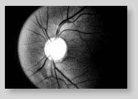
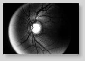
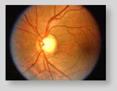
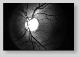
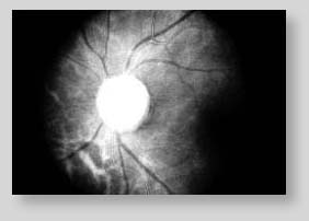
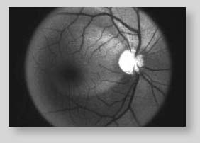
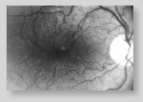
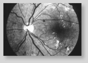
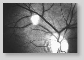
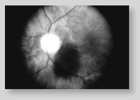
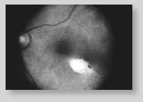
The next series of photographs demonstrates the monochromatic principles and their effect on fundus photographs. The same three primary color filters used for the previous demonstration have been applied to images of the fundus. Various fundus structures become more or less visible with each distinct monochromatic rendition.
Blue light is mostly absorbed by the RPE, retinal blood vessels, choroidal blood and the optic nerve. This provides a very dark background against which the specular reflections and scattering in the anterior layers of the fundus is enhanced. There is little tonal separation between the retinal vessels and the RPE with blue light. Scattering in the ocular media can limit the effectiveness of these wavelengths. The retinal nerve fiber layer, the internal limiting membrane, retinal folds, cysts and epiretinal membranes are examples of scattering structures that may become more evident with short wavelength illumination. Because of excessive scattering at very short wavelengths, blue-green (cyan) filters of 490nm are often employed.
Green light is also absorbed by blood, but is reflected more by the RPE than blue. There is less scatter than with shorter wavelengths, so media opacities have less of a detrimental effect. Green light provides excellent contrast and the best overall view of the fundus. It enhances the visibility of the retinal vasculature, hemorrhages, drusen and exudates. For this reason, green filter "red-free" photos are routinely taken as a baseline in conjunction with fluorescein angiography.
With red light, the RPE becomes a bit more transparent revealing a better view of the choroidal pattern. Overall fundus contrast is greatly reduced with red light. Retinal vessels appear lighter and become less obvious at longer wavelengths. There is greater tonal separation between the retinal veins and arteries; with the arteries appearing lighter than the veins. The optic nerve becomes much lighter and appears almost featureless. Red light is useful for imaging some pigmentary disturbances, choroidal ruptures, choroidal nevi and malignant melanomas.
|
||||||||||||||||||||||||||||||||||||||||||||||||||


