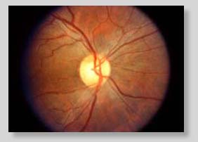
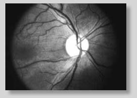
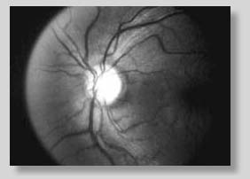
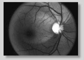
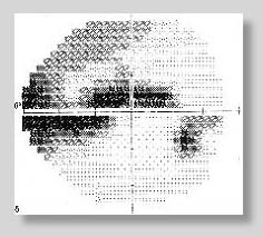
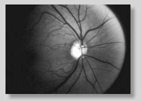
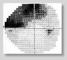
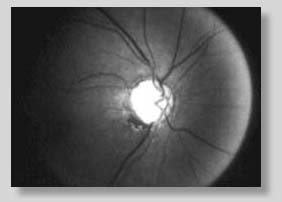
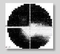
Retinal Nerve Fiber Layer PhotographyTimothy J. Bennett, CRA Monochromatic photography of retinal nerve fiber layer (RNFL) defects may be useful in the detection of early or progressive damage due to glaucoma. A number of studies have shown a correlation between atrophy of the RNFL and visual field loss in glaucoma. Evidence suggests that RNFL photography can detect damage from glaucoma before a visual field loss occurs. With early detection, treatment can be started before extensive damage occurs. RNFL photography may also help predict which glaucoma suspects are at greater risk of future damage. In RNFL photography, we are photographing a semi-transparent subject with a colored background. The retina itself is mostly a transparent structure. The anterior layers scatter light slightly, but it is the base of the retina and the choroid beneath it that provide the reddish-brown color apparent when looking at the fundus. The nerve fiber layer is one of the innermost layers of the retina. It can be seen as a number of faint stripes or striations fanning out from the optic nerve in a radial pattern against the background formed by the retinal pigment epithelium and the underlying blood vessels of the choroid. In a healthy eye, the RNFL gives a cross hatched or slightly blurred appearance to the underlying retinal blood vessels.
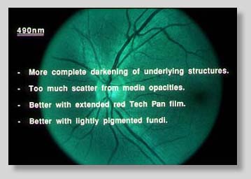
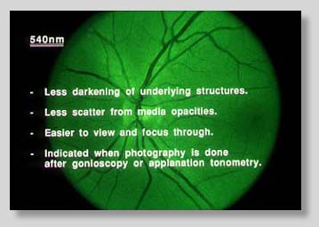
By selectively darkening the background through the use of green or blue-green contrast filters, we can increase the visibility of the RNFL. This darker background enhances the contrast between the nerve fibers; much like a dilated pupil can provide a good background for imaging subtle corneal pathology - another subject that is mostly transparent. In areas of higher concentration of nerve fibers, the light is scattered and some of this light is reflected back through the pupil. The effect of this scattering is enhanced somewhat by the fact that the shorter wavelengths scatter more than longer wavelengths. Due to the normal variation in fundus pigmentation, it is difficult to choose a single contrast filter that works well for all patients. The choice of wavelength often becomes a compromise between the more complete darkening of the underlying pigment with shorter wavelengths and the better penetration of opacities in the ocular media with longer wavelengths. Blue-green filters (470-490nm) produce the best results in patients with lightly pigmented fundi and clear ocular media. Green filters (520-540nm) offer more consistent results in a higher percentage of patients, especially those who cannot be adequately imaged with blue-green.
NERVE FIBER LAYER DEFECTSNerve fiber layer defects can be divided into three different categories - slit, wedge or diffuse. Defects are areas that are darker than normal, although there is a normal light-dark-light pattern to the RNFL. Atrophy of the nerve fibers results in a decrease in thickness of the RNFL. With this decrease in thickness, the normal striped pattern becomes more difficult to observe. There is a decrease in brightness from the RNFL while at the same time the retinal vessel walls can be seen more clearly. Slit defects are dark grooves or stripes that appear wider than the normal striations. They should not be confused with the normal fanning out of bundles as they get further away from the disc. Only those slits that can be traced directly to or near the optic disc margin are considered to be defects. There is usually no corresponding visual field defect. Slit defects are believed to be of less significance than once commonly thought.
Wedge defects are found in the arcuate area and are the easiest to observe. They are usually well defined against adjacent normal areas. Unlike slit defects, there is often a corresponding arcuate visual field defect.
Diffuse atrophy is often the most difficult to detect, yet is considered to be the most significant type of defect. Vessels begin to stand out in bold relief against the more visible mottled appearance of the RPE. Moderate diffuse loss will reveal the more superficial large vessels, while severe diffuse loss will also reveal the small, deeper capillaries. Focal slit or wedge defects can sometimes be found within a larger area of diffuse atrophy. Diffuse atrophy often precedes visual field abnormalities in glaucoma suspects and is of predictive value for further glaucomatous damage.
TECHNIQUE
FILMS FOR RNFL PHOTOGRAPHY
RECIPES
|
||||||||||||||||||||||||||||||||||||||||||||||||||||||||||||||||||||||||||

