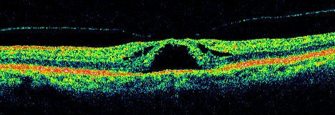
Optical Coherence Tomography
Optical Coherence Tomography (OCT) uses coherent interferometry to construct a cross-sectional view of ocular structures that is accurate to 10 microns. Interferometry is the general technique of superimposing, or "interfering" two or more light waves which creates an output wave that is different from the input waves. In OCT, the ocular structure is illuminated with near-infrared radiation, and the reflected waves are superimposed with waves from a reference mirror. The differences between the reflected waves and the reference waves are used to construct cross-sectional views. OCT is non-invasive, and very accurately documents structural abnormalities within the retina. A detailed on-line course on OCT is planned for this site. This will be the first such course offered by the Ophthalmic Photographers' Society that will provide on-line Continuing Education Credit which can be applied toward the CEC requirement for Certified Retinal Angiographers. The target date for completion of this project is January, 2008.
|

