Processing Angiograms
|
Janice Clifton, CRA COT |
Paul R. Montague, CRA FOPS |
Photographic Illustrations by |

An Ocular fluorescein angiogram is often referred to in ophthalmology circles as either an angiogram or a fluorescein. We will use the term angiogram to mean Ocular Fluorescein Angiogram. Other types of angiography require methods and procedures which differ from those outlined here.
Although digital imaging is quickly becoming the standard method for acquiring and presenting angiographic images, black and white film processing and printing remain an integral part of ophthalmic photography in some areas.
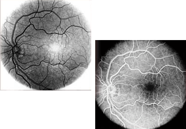
The film on which angiograms are taken must be processed using a photographic developer. This developer creates a negative image on the film. The fluorescein dye, which appears light in the vessels of the ocular fundus, are rendered as dark images on the film against a clear background.
Many ophthalmologists prefer to interpret the fluorescein negatives because some small amount of information may be lost in producing prints, and because simple film processing can be done fairly quickly without the additional steps of printing. Some, however, find it difficult to view the angiogram as a negative and prefer that positive prints be made which render the fluorescein dye as light against a black backround.
