Slit Lamp Biomicrography
|
 |
 |
Retroillumination from the Iris
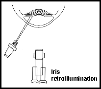
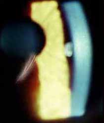
Retroillumination from the iris is created by making a moderately thin slit beam and directing the beam to the iris at a 45 degree angle and keeping the plane of focus on the cornea. The soft reflected beam from the iris will enhance transparent corneal irregularities that are too subtle to be observed with other lighting situations. Details of corneal changes are best observed in the line between light and dark coming from the unfocused iris surface.
 |
 |
Retroillumination from the retina
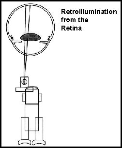
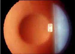
Retroillumination from the retina can be set up by decentering the broad beam and rotating in the beam aperture that most resembles the patient's dilated pupil diameter. (The patient must be dilated for this type of illumination.) The next step is to create a 'half moon' shape by rotating a half aperture position out of the beam path. The decentered 'half moon' shape is then positioned just inside the dilated pupil. The observer must then look for a bright orange reflex that appears when the light is just axial enough to reflect off the patient's retina and glows from behind the lens and cornea. The position for the light and camera should be very close to axial but the retroillumination effect does not appear in both oculars in the binocular biomicroscope. The observer should close one eye and concentrate to see the retro illumination effect in the ocular that the camera shares. The retroillumination effect can be further enhanced or brightened by orienting the patient's eye (have the patient look slightly nasal) to make the optic nerve reflect the beam. This lighting situation enhances the observation of cataracts, corneal scars, transparent cysts, and refractile bodies.
 |
 |
Iris Transillumination
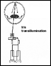
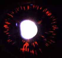
Iris transillumination requires an undilated pupil and is created by making an axial light beam shine into the small pupil and reflect off the retina. The beam aperture should match the pupil size or be smaller than the pupil to avoid iris reflections. The beam direction is again critical to proper alignment and the observer should again use only the ocular shared by the camera to properly align the light beam. This lighting situation is used to illustrate the degree of iris thinning either due to ocular albinism or pigmentary glaucoma. The light transmitted through the iris is the dimmest of all lighting techniques outlined. The flash power must be at its maximum level, or the aperture for the camera should be at its widest opening to make the photograph. Some photographers have more sensitive film on hand just for this occasion. (Typical films for biomicrography are ISO 200 -- a more sensitive emulsion would be ISO 400.)
 |
 |
|
