|
|
 |
Slit Lamp Biomicrography
The Slit Lamp
James P. Gilman, CRA
Director of Ophthalmic Photography
John Moran Eye Center
Salt Lake City, Utah
 The clinical biomicroscope, or "Slit Lamp", is composed of a binocular viewing system that is copivotal with the illuminator to allow various angles of viewing and angles of lighting to the eye. A severe angle of illumination will enhance the surface details and texture by showing a shadowing on the distal edge of the subject. A direct coaxial angle of illumination will accurately show color size and relative position of the subject in relation to other anatomy and will "flatten" the structures that appeared more three dimensional with the texture lighting. The biomicroscope and illuminator are also parfocal or simultaneously focused at the same point in space at all angles. When the slit beam is focused sharply on the cornea, the beam will be in focus through the biomicroscope and will be isocentric or always centered in the viewing field no matter the angle of illumination or viewing.
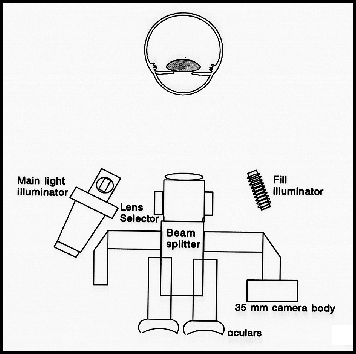
The photographic biomicroscope differs slightly from the clinical biomicroscope by having a beam splitter transmit a percentage (50% or 30%) of light to the viewing oculars and reflect the remainder of the light to the camera. Some photo biomicroscopes have the ability to rotate the beam splitter out of the viewing path to allow 100% of the reflected light to be seen by the examiner. This procedure helps to reveal all the subtle details of the patient's eye without interruption of the light. A safety mechanism on a camera of this type, will prevent the camera shutter from opening without the beam splitter rotated in, which would produce a photograph that would be grossly underexposed. An additional feature of the photographic biomicroscope is the synchronized flash illuminator. The camera accurately records the details simultaneously viewed by the examiner/photographer. Another feature specific to the photo biomicroscope is the fill flash. This allows the photographer to fill in the shadow areas opposite the main illuminator and to provide orientation details of the eye when using a fine slit beam illumination. Without the aid of the fill illuminator the slit beam would give the illusion of floating in the middle of a dark void. This can sometimes be desirable if the photographer is interested in details within the slit beam itself to show corneal edema, particles in the stroma or outline topography with the slit.

The slit beam can take on many configurations from a full round circle to a thin beam of light. The illuminator contains controls for beam aperture size which controls the height of the beam and the beam width. The beam can also be decentered with another control to aim the projected beam off axis for creating a series of indirect lighting situation that will be discussed later.
|
 |
|