|
|
 |
Slit Lamp Biomicrography
Direct Illumination
James P. Gilman, CRA
Director of Ophthalmic Photography
John Moran Eye Center
Salt Lake City, Utah

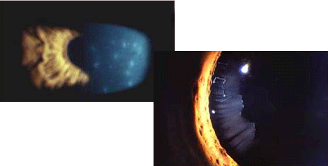
Illumination forms fit into two main categories of direct and indirect lighting. If the lesion is opaque, crystalline, or opalescent, a direct form of illumination delineates the areas of interest better than an indirect form of illumination. If the lesion on the eye is transparent, refractile, and almost invisible, indirect forms of illumination are more successful at enhancing fine details contained in subtle lesions that direct lighting would overpower. First, we will explore the types of Direct Illumination.
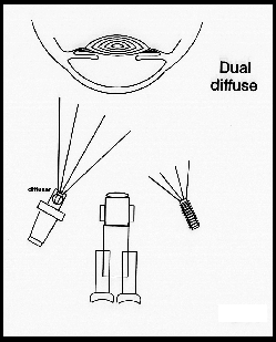
Dual Diffuse
Dual diffuse illumination is achieved when the main light has a diffusion filter in front of the beam and the fill illuminator/flash is on, directed opposite the main light illuminator, to fill in the shadows. This photograph is taken in all cases to allow orientation of the eye being examined, and is typically taken at low magnification (1OX) and at (16X).

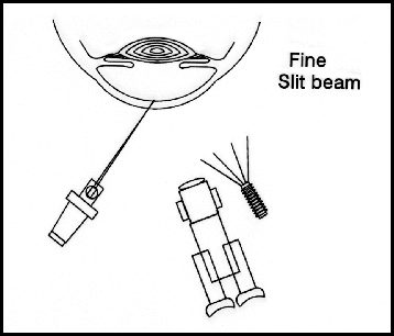
Fine Slit Beam
Fine slit beam illumination is created by removing the diffusing filter from the main light illuminator and reducing the beam width to a fine slit. The fill light is also employed to provide the photograph with surrounding details of the slit's position. The camera or observer oculars should be positioned at approximately 60 degrees to the fine slit beam's direction. Since microscopes will generally have a narrow depth of focus, the 60 degree angle will allow more of the beam depth or optical section to be in focus to the observer and the photograph. The fine slit illumination can show corneal thickening with corneal edema or corneal thinning due to erosion. The fine slit beam can also show topographic information in cases of iris lesions, corneal or conjunctival masses, cataracts and fluid-filled cysts.

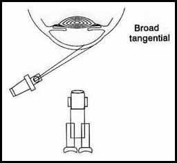
Broad Tangential
Broad tangential Illumination is made possible by increasing the beam width and increasing the angle of incidence for the beam to spread across the cornea. This type of illumination is more effective without the fill illuminator. Broad tangential illumination helps to increase contrast and show varying degrees of texture in the ocular tissue. This lighting is especially helpful for showing slight corneal scarring, pseudoexfoliation of the lens, and small bullae in a bullous keratopathy.

|
 |
|