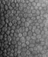
Specular Endothelial MicrographyKirby R. Miller, CRA
The cornea is the clear tissue at the very front of the eye ball. It is somewhat like a watch crystal. It is a very highly specialized tissue. It is optically clear to allow light to pass through the surfaces inside the eye to the retina. Blood vessels don't grow into the cornea. This means that nourishment of this small structure must be unique. The layer of cells on the inside of the eyeball is called the endothelium. This single layer of hexagonal cells is largely responsible for pumping the nourishment into and out of the rest of the cornea. If this cell layer fails, the cornea becomes thick and is no longer clear. Research has shown that we are given a limited number of endothelial cells at birth. If we scratch our skin or the surface of the cornea, the cells grow back to cover that area by duplication. This doesn't happen with the corneal endothelium. If they are damaged due to disease or trauma, they don't grow back. The specular microscope is a special instrument that allows the doctor and photographer to see and record the corneal endothelial cells. Some instruments touch the front or the cornea. Others are non-contact. These instruments take advantage of a bright specular or mirror like image that is created when the angle of the light for photography is at the correct angle. Since the cornea is clear, only a small amount returns to do the photography. It's similar to looking at the inside surface of a drinking glass by changing the angle of light. Contact or applanation specular micrography requires that the patient's cornea be anesthetized with eye drops. The patient's head is then placed into a frame with a chin rest which controls movement. The camera is brought toward the eye and the front element placed in contact with the cornea. The endothelial cells are brought into focus and either photographed on film or recorded on video tape. It is a relatively quick procedure if the patient is very still. Eye movements and tremors can make the procedure difficult since the magnification of the camera has to be relatively high. Non-contact instruments don't touch they eye and are used like the special biomicroscope or slit lamp. Contact instruments have more magnification, but may be impossible to use in difficult patients. Non-contact instruments have much less magnification which may prevent seeing good cell detail. They may be much easier to use in difficult patients and especially children. The images which result from the procedure allow a "cell count". Our corneal endothelial cells decrease as we age. A normal cell count for the population at large would be expressed as 2500 per millimeter squared. The range for normal is much larger, 2000-3200. Ophthalmologists might want a cell count to evaluate a patient prior to cataract surgery. Certain inherited changes cause a premature loss of corneal endothelial cells. A cell count might be ordered for a suspicious change in the appearance or thickness of the cornea. Cell counts have been used to evaluate a wide variety of procedures, primarily those used in cataract surgery. Eye and tissue banks routinely use the specular microscope as one method of evaluating a donor cornea prior to corneal transplant.
|

