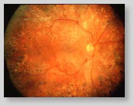
Fundus Imaging OverviewRetinal Fundus PhotographsExcerpt from:  Normal Fundus Photograph Fundus photographs are visual records which document the current ophthalmoscopic appearance of a patient's retina. One picture is worth, in this instance, a thousand words in the physician's notes. They allow the physician to further study a patient's retina, to identify retinal changes on follow-up, or to review a patient's retinal findings with a colleague. Fundus photographs are routinely ordered in a wide variety of ophthalmic conditions. For example, glaucoma (increased pressure in the eye) can damage the optic nerve over time. Using serial photographs, the physician studies subtle changes in the optic nerve and then recommends the appropriate therapy. (Ref: Armaly, MF. Optic cup in normal and glaucomatous eyes. Invest. Ophth. 9(6):425-429)  Glaucoma Fundus photography is also used to document the characteristics of diabetic retinopathy (damage to the retina from diabetes) such as macular edema and microaneurysms. This is because retinal details may be easier to visualize in stereoscopic fundus photographs as opposed to with direct examination. (Ref: Kinyoun, JL, et al, 1992. Ophthalmoscopy versus fundus photographs for detecting and grading diabetic retinopathy. Invest. Opthalmol. & Vis. Science. 33:888-93.)  Diabetic Retinopathy Fundus photography is also used to help interpret fluorescein angiography because certain retinal landmarks visible in fundus photography are not visible on a fluorescein angiogram.
|
