Fundus Imaging OverviewFundus Camera OpticsExcerpt from:
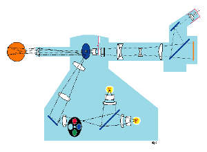
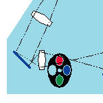
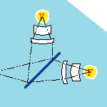
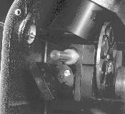
Light generated from either the viewing lamp or the electronic flash is projected through a set of filters and onto a round mirror.
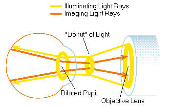
This mirror reflects the light up into a series of lenses which focus the light. A mask on the uppermost lens shapes the light into a doughnut. The doughnut shaped light is reflected onto a round mirror with a central aperture, exits the camera through the objective lens, and proceeds into the eye through the cornea.
Assuming that both the illumination system and the image are correctly aligned and focused, the resulting retinal image exits the cornea through the central, un-illuminated portion of the doughnut.
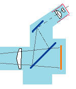
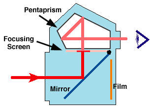
The light continues through the central aperture of the previously described mirror, through the astigmatic correction device and the diopter compensation lenses, and then back to the single lens reflex camera system.
|
|||||||||||||||||||||

