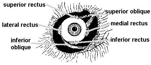
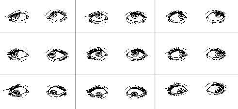
Ocular Motility PhotographyGary E. Miller, CRA Illustrations by Diane Latranyi The term ocular motility refers to the study of the twelve extraocular muscles and their impact on eye movement. Each eye has six muscles, four rectus and two oblique, which, when functioning properly, allow the eyes to work together in a wide range of gaze.  Muscles of the Right Eye Patients are usually tested for eye position and movement during routine eye examination. To observe the action of all twelve muscles, the patient is asked to look in the 9 diagnostic positions of gaze. The patient is instructed to follow a target to various points of gaze while the physician closely monitors their eye movements. Any noted limitation or misalignment of the eyes could indicate muscle weakness or paralysis and warrant further investigation.
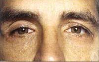
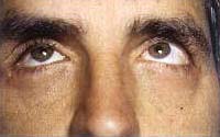
Ocular motility photographs may be requested in order to document any improper muscle action. Patients with strabismus and those diagnosed with cranial nerve palsies are good candidates for photographic documentation. These photographs can be used in reinforcing the physician’s clinical impression, for medical-legal reasons or as a starting point prior to muscle surgery.
|
||||||||||


