|
|
 |
External Eye Photography
Protocols and Magnification
William C. Nyberg, RBP CRA
Scheie Eye Institute
University of Pennsylvania

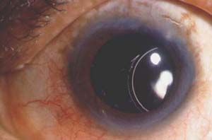
Displaced Intraocular Lens
Protocols
Careful positioning of the patient and the eye is crucial to excellent photography. Images used for scientific documentation deserve to be made with great care and attention to detail. The usual conventions of medical photography for anatomical positioning should be observed. Protocols for camera viewpoint should be developed giving consideration to bone structure and iris position. Magnification protocols should consider telling the pathological story with a series of photos. Typically, intervals of a factor of 2 (each picture taken at twice the magnification of its predecessor) do the job nicely. Once these protocols are developed (usually in conjunction with the physicians who will use the final images), they should be followed religiously, giving the images more scientific utility over time.
Magnification
The face is nicely rendered at about 1/8x (in which the image on the film is 1/8 as large as the subject). Two eyes together, or oblique views of one eye that include substantial lid area are made at 1/4x or 1/3x. Magnifications of 1/2x to 1x are needed to document the eye lids. 3/4x is a personal favorite. The globe is best shown at magnifications from 1x to 2x (the film image being twice as large as the subject). At 2x, a cornea will fill the frame from top to bottom. 1 1/2x will show the entire iris as well as enough sclera above the limbus to include surgical cicatrices. 1 1/4x will show the whole eye including each canthus. 1x pictures are useful for visual establishment as a base for higher magnification detail.
|
 |
|