|
|
 |
External Eye Photography
Techniques
William C. Nyberg, RBP CRA
Scheie Eye Institute
University of Pennsylvania

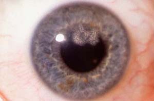
Ghost Vessels on Cornea
Subject Stabilization
Accomplishing photomacrographic imaging of a live patient in a clinical setting is very challenging. The most immediate problem encountered is the natural movement of the patients head. High magnification photography results in very shallow depth of field however it is accomplished, and even small patient movements are enough to seriously disrupt imaging efforts. Verbal encouragement and chin/head rests are the most commonly attempted solution. But having the patient lean his head against a wall, or lie on an examining table and photographing from above will also work.
Focusing
Some 35mm and digital slingle-lens reflex cameras have interchangeable focusing screens. Some of these screens are designed to work with high magnification images and dim lighting conditions, and are definitely worthwhile. Perhaps the most valuable aid to focusing an external camera on lesions in the anterior chamber of the eye is to do slit lamp photography before attempting external photography. The education obtained during the slit lamp exam will allow the photographer to approach the viewfinder of the single lens reflex camera with confidence, as he/she will know the exact location and appearance of the lesion. Focusing the camera should be done by setting a preselected magnification and then moving the entire camera/lens apparatus until the image in the viewfinder is clear. This technique maintains consistent magnification. The usual practice of rotating the focusing collar on the lens changes the magnification of the image. This is perfectly acceptable in normal photography, but not in scientific photography.
|
 |
|