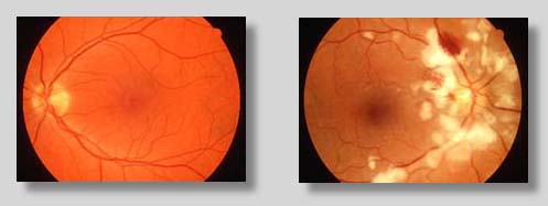
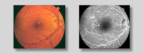
Ophthalmic PhotographyOphthalmic photography is a highly specialized form of medical imaging dedicated to the study and treatment of disorders of the eye. It covers a very broad scope of photographic services incorporating many aspects of commercial and medical photography. But it is through the use of highly specialized equipment used to document parts of the eye like the cornea, iris, and retina, that ophthalmic photography takes on it's true identity. The retina is the "film" of the eye. Images passing through the clear structures of the cornea and lens are focused there to give us our view of the world. Special instruments called fundus cameras, when used by skilled photographers, can document the condition of this miraculous anatomical structure.
When fundus photography is performed after the injection of a fluorescent dye into the bloodstream via a vein in the patient's arm, the procedure is called Fluorescein Angiography. With special colored filters, only the dye is photographed as it travels through the vessels in the retina. These studies, performed by ophthalmic photographers and interpreted by ophthalmologists, are used in differentiating one retinal disease from another and in determining appropriate courses of treatment.
A horizontally mounted microscope, coupled with special illumination devices is used to photograph the cornea. This photo slitlamp produces high magnification views of disorders that would be impossible to observe with the naked eye. These and many other techniques employed by ophthalmic photographers to aid in the diagnoses and treatment of disorders of the eye are described here. Nearly all ophthalmic photographers perform fundus photography and fluorescein angiography. Many of the other skills are required in many settings. |